An Ophthalmoscope Is Used to Determine Which of the Following
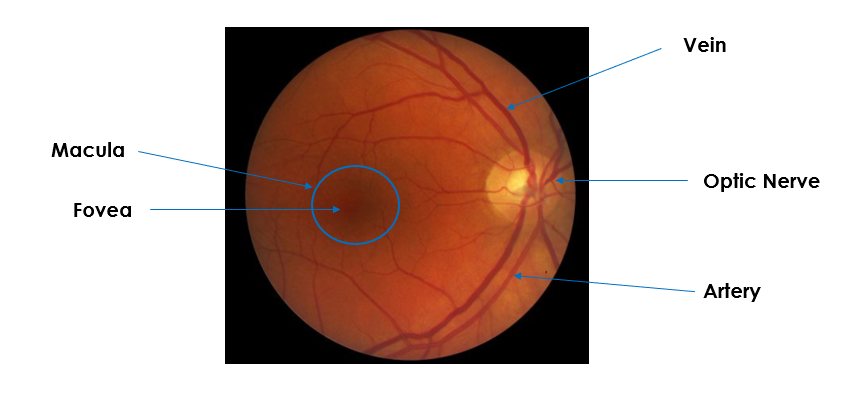
What am I looking for. The ophthalmoscope also known as a fundoscope is a tool used in medicine to examine the interior of the eye including the retina fovea choroid macula optic disc and blood.

Moran Core How To Use The Direct Ophthalmoscope
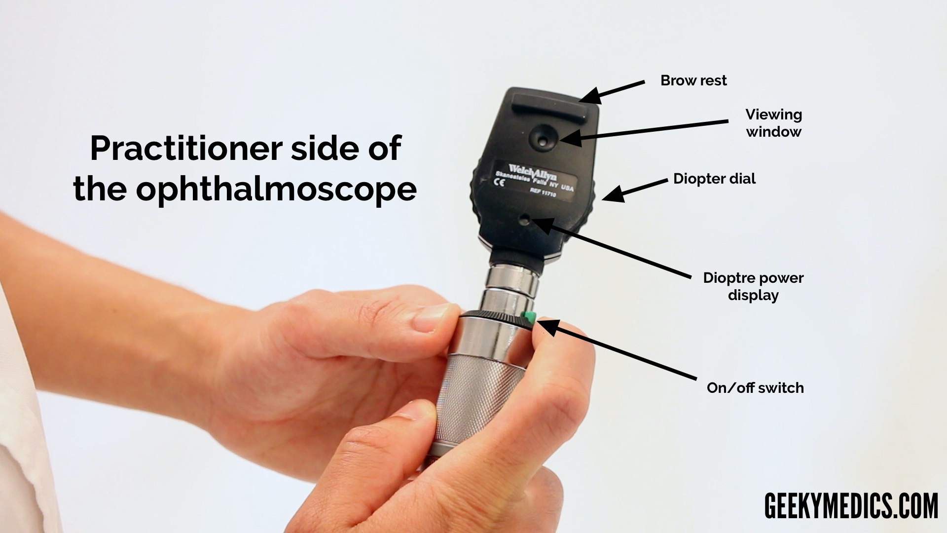
Which parts of the ophthalmoscope are present on the front end of the ophthalmoscope head.
. An ophthalmoscope is an instrument used to examine the retina. There are two main types of. These include hyperemia and elevation of the optic disc with obscuration of the.
12126129 A 15 minute indirect ophthalmoscope. Which assessment technique should the nurse use to determine the body temperature of a patient. Which aperture would be used to assess the eyes of a patient with undilated pupils C small at the conclusion of the examination the examiner should D summarize findings to the patient When the practitioner enters the examining room the infant patient is asleep.
Most ophthalmologists have deserted the direct ophthalmoscope for a different device an indirect ophthalmoscope which provides a better view of the peripheral retina and assists in the discovery of retinal holes etc. If youve ever been for an eye test or visited an ophthalmologist theres a good chance they would have taken a look at your retina with an ophthalmoscope. Funduscopy with a direct ophthalmoscope or indirect slit-lamp ophthalmoscope remains the easiest and most sensitive means of detecting papilledema.
Your eye doctor can use ophthalmoscopy to screen for eye diseases and conditions that can affect blood vessels. Traditionally part of almost every eye exam ophthalmoscopes can identify healthy structures within the eyeball and easily help your eye doctor see symptoms or indicators of diseases of the eye. S face move an object inward from the periphery.
Damage to your optic nerve. In addition some digital ophthalmoscope feature backlit screens to make them easier to see in bright light. Touching the patients skin with the dorsal side of the hands and fingers.
Detect foreign bodies in the cornea b. The ophthalmoscope selection should be guided by a number of factors. In its full-blown form papilledema has all the manifestations of axoplasmic constipation and peripapillary venous obstruction Figure 70-1.
Pediatricians and general practitioners may also include ophthalmoscopy in routine physical exams. Others have auto-shutoff features that turn them off after a few minutes of inactivity. All of the above.
Advantages of direct ophthalmoscopy have traditionally included 1 a 15 magnified view of the posterior pole that facilitates appreciation of small dynamic changes of the ocular fundus such as venous pulsations and circulatory changes. Ophthalmoscopy also called fundoscopy is an exam your doctor optometrist or ophthalmologist uses to. Sit facing the client and while look directly at the client.
Prime among these is the intended clinical role. Turner in Retina Fifth Edition 2013 Ophthalmoscope and fundus camera exposure. Verify doubtful papillary action c.
An ophthalmoscope is a piece of equipment utilised by ophthalmologists that are used to inspect the internal structure of your eyes containing the retina. Ophthalmoscopy also called funduscopy is a test that allows a health professional to see inside the fundus of the eye and other structures using an ophthalmoscope. You will either lie.
The lenses of the ophthalmoscope can be used to focus variously the apex and base of any intraocular mass and thus helps determine its height in dioptres. An ophthalmoscope is about the size of a flashlight. An expected part of every eye exam ophthalmoscopes are.
With a manual range ophthalmoscope it can be difficult to determine the approximate range of electrical current youre testing. Used to examine contour abnormalities of the cornea lens and retina. The exam involves the use of special lenses and bright direct light to provide a better.
They are used all over the world and are an essential piece of apparatus for all who wish to study the intricate biology of the eye. Ophthalmoscopy also called fundoscopy or a fundoscopic exam is a common procedure performed by an eye doctor. Retinal tear or detachment.
It is done as part of an eye examination and may be done as part of a routine physical examination. The ophthalmoscope can also be used for examining the anterior part of the eye by turning the lens dial to 10. Reviewed by Brian Boxer Wachler MD.
Used to make rough. Shine the ophthalmoscope light into the patients pupil at arms length and observe the red reflex. Some ophthalmoscopes have this feature that can be used to observe corneal abrasions and ulcers after fluorescein staining.
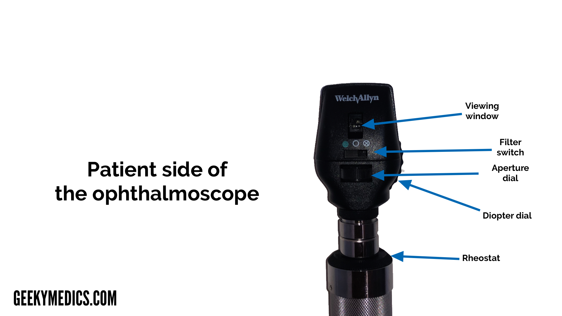
2 wide availability and portabilitythe direct ophthalmoscope is easily carried in the pocket of a clinicians coat and. Mirror window Viewing aperture. You will be seated in a darkened room.
An ophthalmoscope is particularly useful for examining the structures of the retinathe light sensitive area at the back of the eye responsible for processing images. Use an ophthalmoscope to watch the clients pupil constrict when a strong light is shown on it. Red reflex anterior segment disc vessels and lastly macula see box.
Detect lens opacities d. It has a light and different tiny lenses that allow the provider to view the back of the eyeball. The ophthalmoscope has 5 apertures.
The pupil is a hole through. How to select an appropriate ophthalmoscope. The ophthalmoscope was invented by Helmholtz in 1850.
The health care provider performs this exam by shining a beam of light through the pupil using an instrument called an ophthalmoscope. Indirect ophthalmoscopes and fundus cameras produce dazzling exposures and transient afterimages but there is no evidence that they cause photochemical or thermal retinal injury in routine clinical use. It is crucial in determining the health of the retina optic disc and vitreous humor.
Using the ophthalmoscope light as a pen light briefly examine the external features of the eye including lashes lid margins conjunctiva sclera iris and pupil shape size and reactivity. For a century it was the only way to view the fundus of the eye. Medically Reviewed by Whitney Seltman OD on May 08 2021.
14033332238 Confrontation technique during a vision exam is used to determine peripheral vision so D allows the client. The ophthalmoscope can be used to.
Fundoscopy Ophthalmoscopy Osce Guide Geeky Medics
Moran Core How To Use The Direct Ophthalmoscope
Fundoscopy Ophthalmoscopy Osce Guide Geeky Medics
No comments for "An Ophthalmoscope Is Used to Determine Which of the Following"
Post a Comment